Prague Node – Advanced Light and Electron Microscopy Multi Modal Multi Sited Node
About Prague Node
Locations: Prague and České Budějovice
Number of Core Facilities: 7
Technologies & expertise:
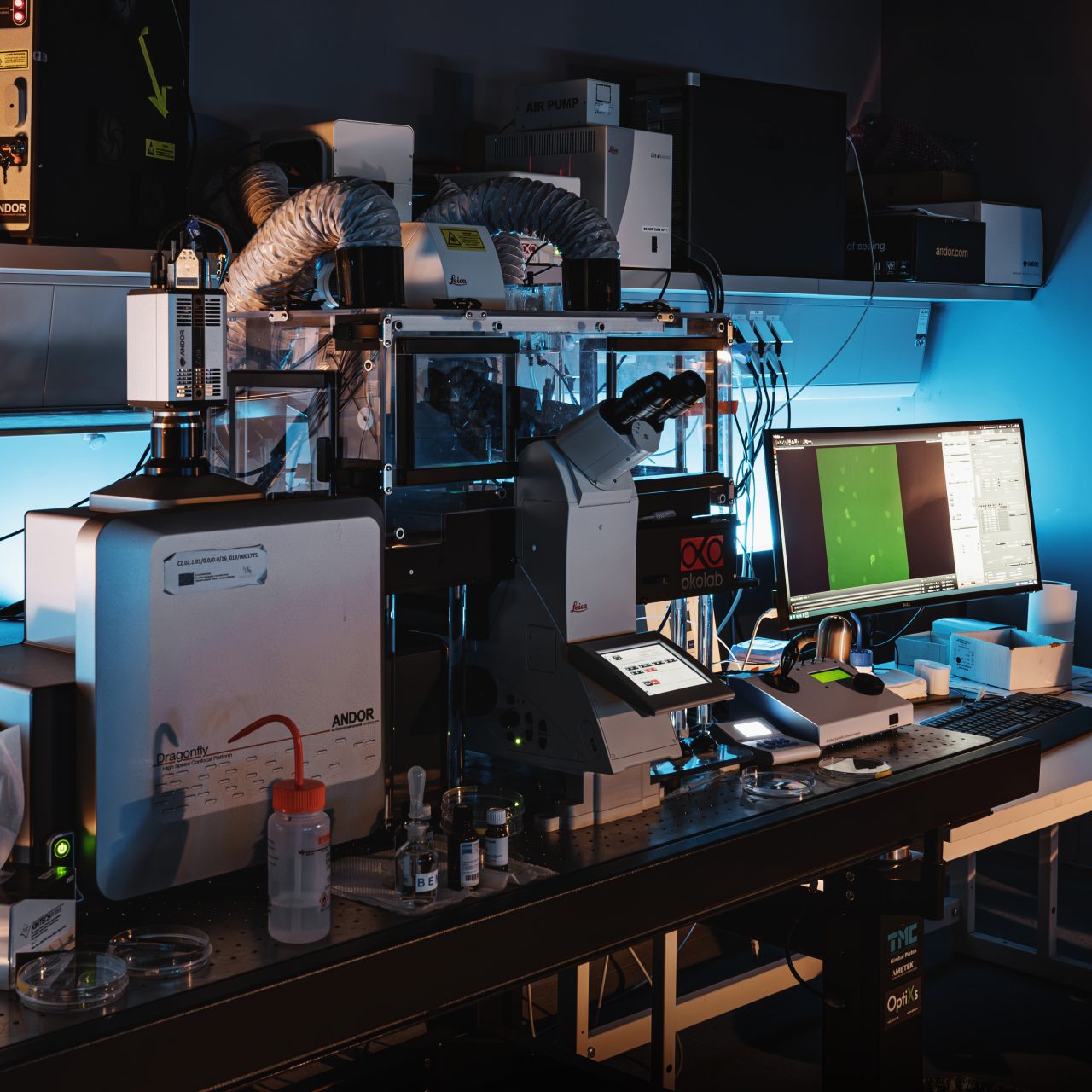
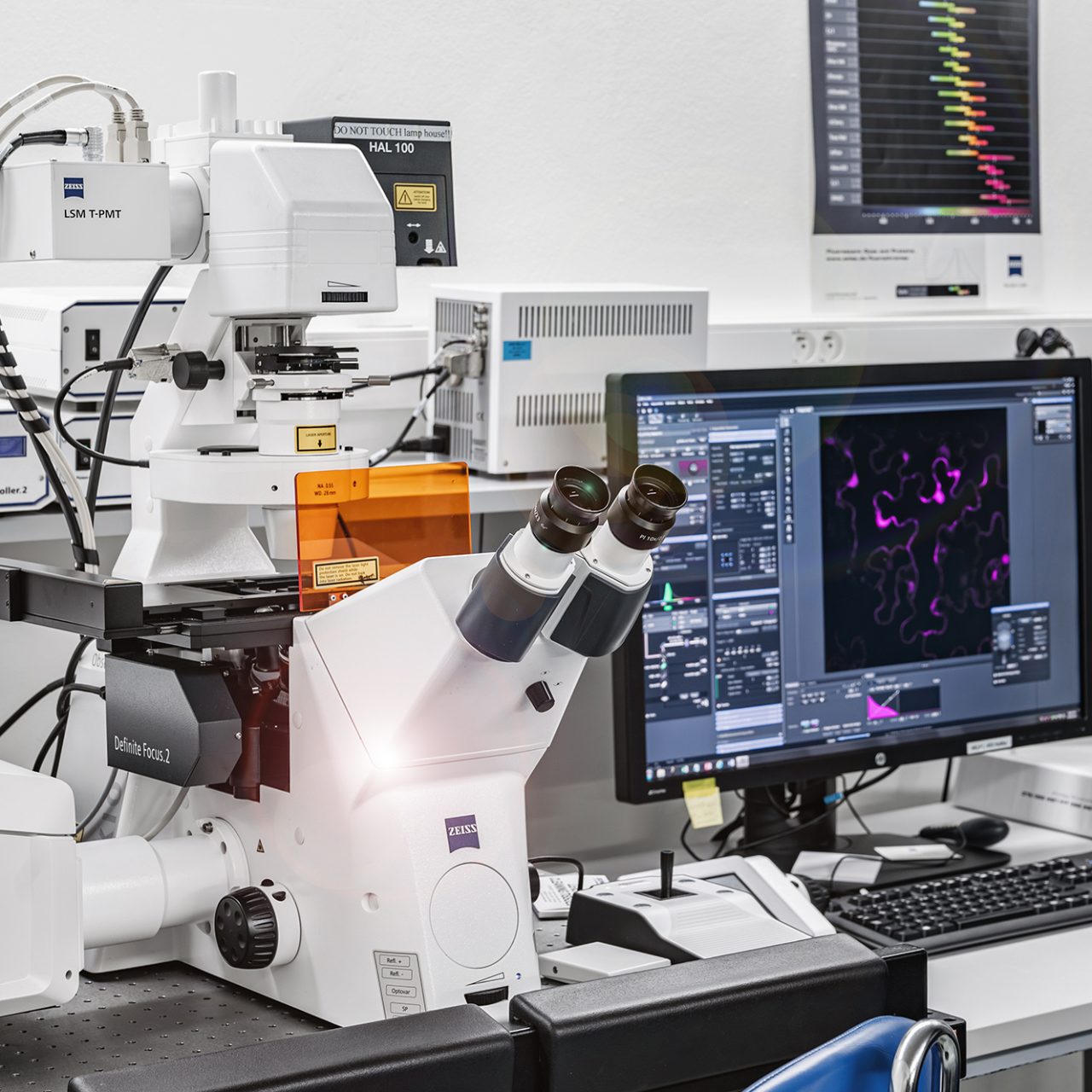
Light microscopy: Multi-functional point and spinning disc confocal microscopes, light-sheet and intravital systems, high-end multi-photon and super-resolution systems
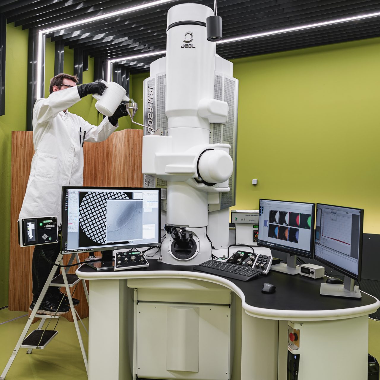
Electron microscopy: Biological sample preparation, ultrastructural imaging techniques (including cryo techniques), volume EM methods (electron tomography, FIB-SEM, SBF-SEM, AT), EDS elemental analysis, CLEM (Correlative Light and Electron Microscopy)
Specialties and expertise of the Node
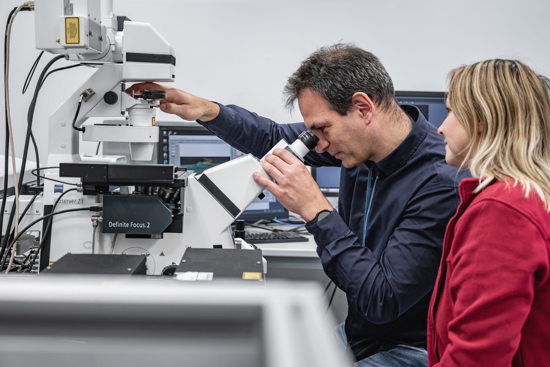
The main strength is the wide and deep expertise covering most of light and electron microscopy biological applications, allowed by the complementary nature of collaborating core facilities and more than 25 FTE of expert staff operating more than 50 advanced microscopy systems. In super-resolution imaging all main approaches are covered, including STED, SIM and SMLM, by multiple commercial systems from different manufacturers.
Information about molecular dynamics and interactions can be obtained by functional imaging, particularly from spatial-temporal correlation analysis (point, line and image F(C)CS), FRAP, photoactivation, FRET, FLIM or PLIM.
Label-free imaging profits from non-linear processes induced by femtosecond NIR lasers and offers methods like SHG, THG, autofluorescence FLIM including metabolic imaging and CARS not only for lipid droplets visualization. Innovative low-toxicity label-free imaging is offered by quantitative phase imaging (QPI).
Plants growing in their natural gravity conditions can be directly visualized on the vertical microscope stage of CZ LSM880 with Airyscan. Fast 3D acquisitions are covered by spinning disc and light-sheet systems.
In the light intravital microscopy (IVM), the node offers a complete package of services: advanced 2-photon imaging systems, infrastructure for the care and management of small rodents and assistance with surgeries.
In electron microscopy the Node offers complete workflow from sample preparation (room temperature or cryo- methods, immunolocalization) through various imaging modalities (TEM, SEM, SBF-SEM, FIB-SEM, array tomography, STEM-EDS elemental analysis) to data analysis (clustering and colocalization in immunolabeling, 3D visualizations). Targeted CLEM workflows allow 3D ultrastructural imaging of rare structures.
Data analysis ranges from commercial software packages (Imaris, Huygens, Amira, NIS-Elements and many more) via custom modified routines for image processing and analysis (for example image reconstruction, registration and semi-automatic and AI assisted segmentations, volume reconstruction and 3D visualization, tracking, colocalization analysis, mathematical modeling and analysis of photokinetic experiments, and more) to new software and modules development in the field of stereology and spatial statistics, FLIM and FCS methods.
Instrument highlights
- Leica TCS SP8 STED 3X and Abberior Instruments Easy 3D STED
- DeltaVision OMX™ V4
- Zeiss Elyra 7
- Leica STELLARIS 8 FALCON
- Leica SP8 AOBS WLL MP
- Bruker Ultima IntraVital
- Femtonics FEMTO3D Atlas
- Carl Zeiss LSM 880 NLO
- Nikon CSU-W1 with FRAP
- Andor Dragonfly 503
- Olympus SpinSR10
- Nikon iLas 2 ring-TIRF with FRAP
- Carl Zeiss Lightsheet Z.1
- Vertical-stage plant-optimized Carl Zeiss LSM880 with Airyscan
- Akoya PhenoCycler-Fusion
- TESCAN Q-PHASE
- Jeol JEM-F200 “F2” with STEM and EDS
- Thermo-Fisher Helios NanoLab 660 G3
- Thermo Fisher UC Apreo VolumeScope SEM
- JEOL JEM-2100F with GATAN K2 Summit direct detector
- TESCAN Amber Cryo with nanomanipulator
- Leica THUNDER Imager EM
- SP8 Cryo CLEM

Core facilities
- Electron Microscopy Core Facility, Institute of Molecular Genetics of the Czech Academy of Sciences
- Light Microscopy Core Facility, Institute of Molecular Genetics of the Czech Academy of Sciences
- BioImaging Facility, Institute of Physiology of the Czech Academy of Sciences
- Microscopy Service Centre, Institute of Experimental Medicine of the Czech Academy of Sciences
- Imaging Methods Core Facility, BIOCEV, Charles University
- Microscopy Facility, Institute of Experimental Botany of the Czech Academy of Sciences
- Laboratory of Electron Microscopy, Biology Centre of the Czech Academy of Sciences
Prague Node hub

Deputy coordinator
Pavel Hozák
Strategic representative
Aleš Benda

Project manager
Daniela Klimešová


