Newsletter Winter 2022
Focus on technologies
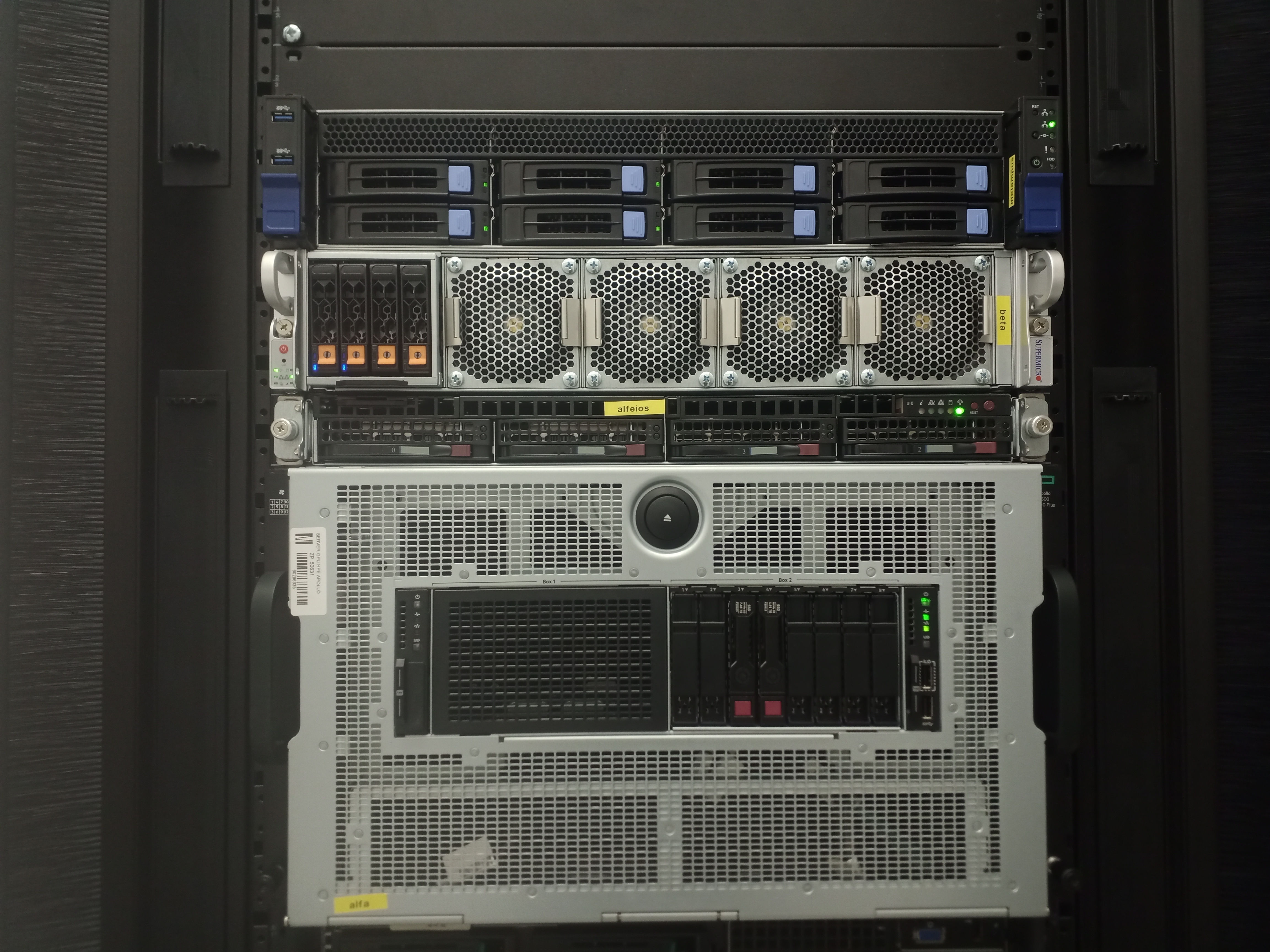
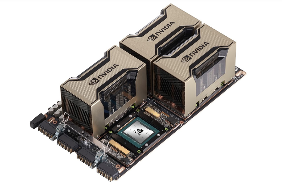
Powerful servers ready for hosting artificial intelligence at Masaryk university
Author: D. Svoboda
The two powerful computational servers are currently provided by the Centre for Biomedical Image Analysis at Masaryk university. Each server is equipped with 0.5TB RAM and four NVidia A100 graphic cards that in total have 240GB memory for highly parallel and accelerated computing.
Our core facility is proud of a high user comfort. The servers are located in the university data center which guarantees the non-stop service and avoidance of blackouts. The broadband connection to the internet, the graphical user interface for each user, and full interactive control without the necessity of creating the batches and scheduling make the use of our service as easy and comfortable as possible. The stable OS Linux Ubuntu 20.04 is installed on both servers. The users of MS Windows operating system are nevertheless not expelled as we can prepare MS Windows desktop environment as well.
A detailed description of the servers and services provided by the Centre of Biomedical Image Analysis can be found at https://cbia.fi.muni.cz/services/. The services are provided to both internal and external users. In case of interest, please contact the support team at cbia-supp@fi.muni.cz.
Highlights of user results
Quantitative MRI of the cervical spinal cord
Author: J. Valošek
A multi-year effort of an international research group led by prof. Josef Bednařík has recently resulted in several journal articles elaborating on the usage of advanced magnetic resonance imaging (MRI) techniques in patients with degenerative cervical spinal cord compression.
The MRI protocol for 3 Tesla human MRI scanners optimized by the group allowed the acquisition of high-resolution diffusion MRI, MR spectroscopy, and structural MRI data. Data were collected on a large cohort of patients with different stages of degenerative cervical spinal cord compression and were related to healthy volunteers. Acquired images allowed to reveal micro- and macro-structural damage of the spinal cord in patients with compression and its relationship to the clinically used measures. Findings serve as a starting point for the follow-up longitudinal study with the aim to identify the predictive MRI-based biomarkers of spinal cord damage.
Moreover, our group participated in an international project focused on the development of reproducible MRI protocol for human spinal cord imaging. The protocol was published in the Nature Protocol journal and is widely adopted by researchers all around the world. Over forty centers worldwide have already acquired MRI data with this protocol and published them freely online in line with open-access practices.

Past & upcoming events
IMAGING PRINCIPLES OF LIFE 2022
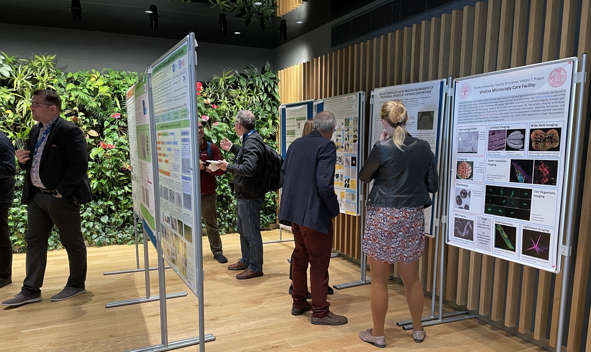
The annual Czech-BioImaging scientific conference Imaging Principles of Life 2022 took place on 4 - 5 October 2022 in Hustopeče, the heart of vineyards and almond trees.
The conference is traditionally dedicated primarily to the Czech-BioImaging users and scientists interested in biological and medical imaging and this year was not an exception. Five scientific sessions were mixed of lectures from fields of electron microscopy, light microscopy and medical imaging, some including an industrial presentation introducing advances in manufacturing. To express in numbers, 31 high-quality scientific talks were given in total. Five of the talks related to the industrial sector and were either given by the company representatives or associated users. The audience reached around 100 participants, which is considered a very satisfactory number for the conference of its kind.
Although all of the lectures presented research of very high-quality, three of them stood out in particular. Two international lecturers accepted the invitation to join the conference and give an invited lecture – Julia Fernandez-Rodriguez, University Gothenburg, Sweden introduced the “Centre for Cellular Imaging – a Research Infrastructure for Correlated Multimodal Imaging” in her talk and Claus Lamm, University of Vienna, Austria focused on “Novel multi-method neuroimaging approaches to unravel the complexities of the human social brain“. Highly acclaimed was the lecture given by Ivan Rektor, Masaryk University, Brno titled “The lifelong impact of extreme stress on the human brain: a three-generation study of Holocaust survivors”.
An integral part of the conference was the poster session in which around 20 scientific posters were displayed. The conference was supported by the Ministry of Education, Youth and Sports of the Czech Republic and commercial sponsors: Abberior Instruments GmbH, Carl Zeiss spol. s.r.o., HPST s.r.o., Měřicí technika Morava s.r.o. and Pragolab s.r.o., who participated in a commercial exhibition introducing their newest microscopic equipment and offering services to the participants.
We are pleased to announce that we are already making arrangements to hold the conference in October 2023.
All
of you are kindly invited to join us. You can expect more information to appear soon on our website
czech-bioimaging.cz, or in your mailbox.

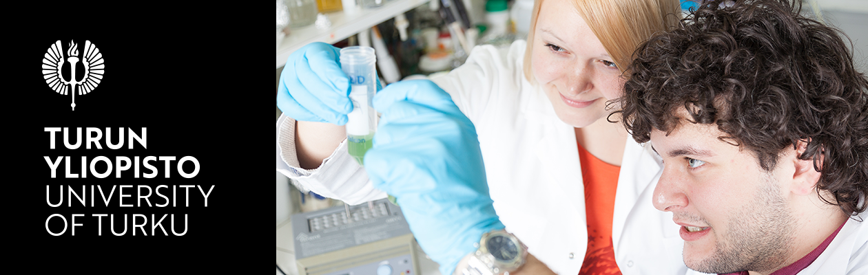
International Master´s Degree Programme in Biomedical Imaging
Do you want to study in a stimulating research environment in a country with beautiful nature and high-tech innovations?
Are you interested in studying Biomedical Imaging in the laboratories where Stefan Hell did his Nobel prize-awarded research with super-resolution?
If your answer is YES, please read this message carefully!
International Master´s programme in Biomedical Imaging is a two-year (120 ECTS) programme jointly administrated by the Faculty of Science and Engineering at Åbo Akademi University, Turku, Finland, and the Faculty of Medicine at the University of Turku (Masters programme in Biomedical Sciences). Close cooperators are the national Turku PET Centre, Turku Center for Disease Modeling, Turku Centre for Biotechnology, and the Turku University Hospital.
Programme is aimed at students with a B.Sc. degree in the Life Sciences or applicable areas of biomedical sciences, physics, chemistry, or engineering. The interdisciplinary curriculum provides the graduates with a broad spectrum of the most recent knowledge in biomedical imaging related to many application areas in cell biology, medicine, and nanotechnology. One of the great examples of our diverse success is the 2014 Nobel Prize in Chemistry shared by Prof. Stefan W. Hell, who has made his original publications on STED microscopy in Turku.
The programme is tuition free for European students and non-EU/EEA students the tuition fee is 12 000€ / year. Scholarships are available!
The language of instruction is English. Book and take the language test well in advance before the application period!
Please find more info in:
- http://www.bioimaging.fi/programme/
- Biomedical Imaging | Åbo Akademi University (abo.fi)
- Master's Degree Programme in Biomedical Sciences: Biomedical Imaging | University of Turku (utu.fi)
- Studying in Finland: http://www.studyinfinland.fi/
- https://www.turku.fi/en/study-turku
Application period: 4-18 January 2023.
We are more than happy to answer if you have any questions about the Master´s Degree programme in Biomedical Imaging! Please contact us: bima-office@bioimaging.fi
OTHER ACTIVITIES
ICRI 2022 participation
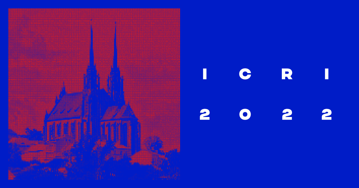
In late 2022, continued to actively participate in national initiatives and other related events.
We are proud to have been a part of the International Conference on Research Infrastructures (ICRI) 2022 held on 19-21 in Brno with the Imaging Principles of Life being the ICRI Satellite event.
Euro-BioImaging membership
Czech-BioImaging also continued its cooperation with its European colleagues, especially through the Euro-BioImaging infrastructure.
Euro-BioImaging ERIC was one of the 3 partners, who joined a new, promising collaborative agreement among top-class life science research infrastructures during ICRI 2022. The trilateral Memorandum of Understanding was signed by 2 other parties - Instruct-ERIC and EU-OPENSCREEN ERIC. The initiative is designed to benefit the life science research community by building common pipelines for user access, and joining forces in training, external communication, FAIR data management, and funding opportunities.
PF 2023
Dear colleagues and friends,
this holiday season, we would like to send you our warmest wishes and thank you for yet another wonderful year of cooperation. We are looking forward to the new era Czech-BioImaging will enter in 2023! Wishing you and your family a happy and healthy holidays and cheers to the New Year, with our very best regards,
Pavel Hozak & the Czech BioImaging team
Useful links
Czech-bioimaging Scientific Conference 2022
Czech-bioimaging – pro veřejnost
The National Infrastructure for Biological and Medical Imaging,
Czech-BioImaging, is supported by the Ministry of Education, Youth and
Sports of the Czech Republic (project No. LM2018129) and by European
Regional Development Fund (project No.
CZ.02.1.01/0.0/0.0/18_046/0016045).
