Newsletter Summer 2023
Focus on technologies
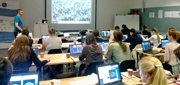
Utilizing Deep Learning Approaches for Daily Image Segmentation Practice at LMCF
Author: Martin Čapek, Ph.D. | LMCF-IMG |
Nowadays, deep learning (DL) approaches for image processing and analysis are being increasingly utilized in real-world and routine practices. These tasks include, for example, deconvolution, denoising, image-to-image translation, object detection, and segmentation. The Light Microscopy Core Facility at the Institute of Molecular Genetics has effectively incorporated several of these techniques in recent years. Here, we will showcase practical examples that demonstrate the application of DL to image segmentation.
One of the most popular and freely available DL projects is StarDist, which we utilized for the segmentation of nuclear protein c-Fos expression spots in images of mouse brain physical slices acquired through widefield transmission microscopy (Fig. 1). The StarDist network was trained using several subpictures extracted from the physical slices, and subsequently applied to the entire images. Fig. 1 shows the original data with color-coded labels using a "3-3-2 RGB" look-up table in ImageJ/Fiji, resulting in various colors in the picture. Fig. 1A focuses on a zoomed-in area with spots from the yellow rectangle in the upper left portion of the slice, while Fig. 1B depicts a corresponding zoomed-in area showcasing the identified labels. Fig. 1C represents a heatmap illustrating the distribution of spots in the brain, with warmer colors indicating a higher spot density. The production of the heatmap involved a custom-written plugin for ImageJ/Fiji.
(Fig. 1 is courtesy of Dr. Helena Janíčková, Laboratory of Neurochemistry, Institute of Physiology of the Czech Academy of Sciences, Prague)
Another valuable DL method is Cellpose. Fig. 2A showcases an optical section of DAPI stained nuclei in a mouse Langerhans islet captured using a Z.1 Zeiss Lightsheet microscope, while Fig. 2B exhibits the section with segmented nuclei labeled. Fig. 2C provides a visualization of the complete 3D stack with segmented nuclei. The objective of this segmentation is to count the number of nuclei in Langerhans islets and compare them to the expected DNA content in the islets.
(Fig. 2 is courtesy of Dr. David Habart, Laboratory for Pancreatic Islets, Institute for Clinical and Experimental Medicine (IKEM), Prague)
Omnipose is a project based on Cellpose and focuses on the segmentation of bacterial species. We utilized it for the segmentation of bacteria in phase data (Fig. 3 A, B) and in Andor Dragonfly spinning disk fluorescence (mCherry) time-lapse images captured over 80 minutes in widefield mode, with four time points shown (Fig. 3 C, D). The left side represents the original data, while the right side shows the segmented data. The purpose of this segmentation is to investigate phenomena associated with bacterial division.
(Fig. 3 is courtesy of Dr. Ondřej Černý, Laboratory of Infection Biology, Institute of Microbiology of the Czech Academy of Sciences, Prague)
MitoSegNet is a suite of tools designed for segmenting and analyzing mitochondrial morphology. In this study, we retrained the model using our own collection of mitochondrial images. Fig. 4A and C present the mitochondrial networks of the control and treated samples, respectively, while Fig. 4B and D display their respective segmentations. The tables in Fig. 4E, generated using a tool provided by MitoSegNet, showcase selected morphological parameters. These tables demonstrate that the morphological parameters are not clustered in the case of the control data, but exhibit strong clustering in the peroxide-treated data.
(Fig. 4 is courtesy of Dr. Jana Vojtová, Laboratory of Regulation of Gene Expression, Institute of Microbiology of the Czech Academy of Sciences, Prague)
We would like to emphasize that the aforementioned examples illustrate challenging segmentation scenarios: Fig. 1 depicts data with an inhomogeneous background and varying spot intensities. Fig. 2 represents blurred 3D data where objects may be touching. Fig. 3 showcases closely touching species. Fig. 4 demonstrates mitochondria with different intensities and thicknesses. Straight and elongated fragments are expected to mostly connect, while round fragments tend to separate.
If you require assistance with your image data analysis and processing tasks, please feel free to contact us or visit our facility.

Highlights of user results

Unveiling the distinct patterns of dynamism among large-scale brain networks that support cooperative and competitive interactions
Author: Kristína Czekóová, Ph.D. | CEITEC, MUNI |
Humans spend most of their lives interacting with other people in various social contexts. Each social exchange is highly complex, requiring us to constantly process multiple social cues from our interaction partner(s) to infer their motivations, intentions and emotions, and adjust our behaviour accordingly.
This is complicated by the reciprocal nature of social interactions, whereby our interaction partners simultaneously modify their behaviour in response to our own. To support interpersonal behaviour, then, the brain must be capable of deploying and switching flexibly between the different functional brain networks that support these complex mental operations. Investigating the neural mechanisms coordinating social interaction therefore requires experimental paradigms and sophisticated equipment capable of capturing brain function from all interactants simultaneously during real social exchanges. In the Central-European region, the acquisition of such complex data is possible thanks to the Multimodal and Functional Imaging Laboratory (MAFIL) at CEITEC MU that is equipped with two adjacent 3T MRI Siemens Prisma scanners and employs biomedical engineers capable of implementing the advanced techniques required for analysing these data. In a close cooperation with MAFIL core facility, Dr. Daniel Shaw and his team recently investigated how large-scale brain networks communicate with one another during cooperative and competitive social interactions. Employing a novel combination of sophisticated analytical techniques revealed that these distinct types of social exchange elicited systematic patterns of dynamical integration and segregation among several core brain networks. Importantly, it was possible to predict with above-chance accuracy the type of interaction taking place in an independent sample from just these patterns of inter-network communication. These findings provide the first direct evidence of systematic communication among large-scale brain networks supporting distinct types of social interaction, which will facilitate the development of neurocognitive models of interactive behaviour and the identification of biomarkers for the interpersonal dysfunction common to many neurological and psychiatric disorders.
The interacting brain: dynamic functional connectivity among canonical brain networks dissociates
cooperative from competitive social interactions.
Shaw, D. J., Czekóová, K., Mareček, R., Špiláková, B. H., & Brázdil, M. (2023)
NeuroImage 269, 119933
doi:
10.1016/j.neuroimage.2023.119933

upcoming event

UPCOMING Educational activities
(summer - autumn)
Fundamentals of light microscopy
Course | June 13-15, 2023 | CEITEC MU, Brno
Entry-level super-resolution microscopy
Course | June 14-15, 2023 | IEM, Prague
Biological Specimens in Electron Microscopes
Course | June 19-23, 2023 | LEM BC, České Budějovice
Image analysis and data processing in superresolution microscopy
Course | August 21-25, 2023 | Viničná MCF, Prague
ImgLib2 and BigDataViewer ecosystem workshop - efficient handling of large bioimaging data with Fiji
Course | Summer 2023 | CEITEC MU, Brno
Advanced Methods in Biomedical Image Analysis
Course | September 10-16, 2023 | FI MU, Brno
Advanced methods of high-resolution SEM
Course | September 25-27, 2023 | VMCF, Prague
Single molecule microscopy and manipulation
Course | October 9-13, 2023 | UK, BIOCEV, Vestec
Microscopy methods in biomedicine
Course | October 16-20, 2023 | IMG, Prague
Multi-modal light microscopy imaging in plant research
Course | October 18-19, 2023 | IEB, Prague
View all courses
Useful links
The National Infrastructure for Biological and Medical Imaging,
Czech-BioImaging, is supported by the Ministry of Education, Youth and
Sports of the Czech Republic (project No. LM2023050) and by European
Regional Development Fund (project No.
CZ.02.1.01/0.0/0.0/18_046/0016045).
Supported by:
