Newsletter Spring 2022
Focus on technologies
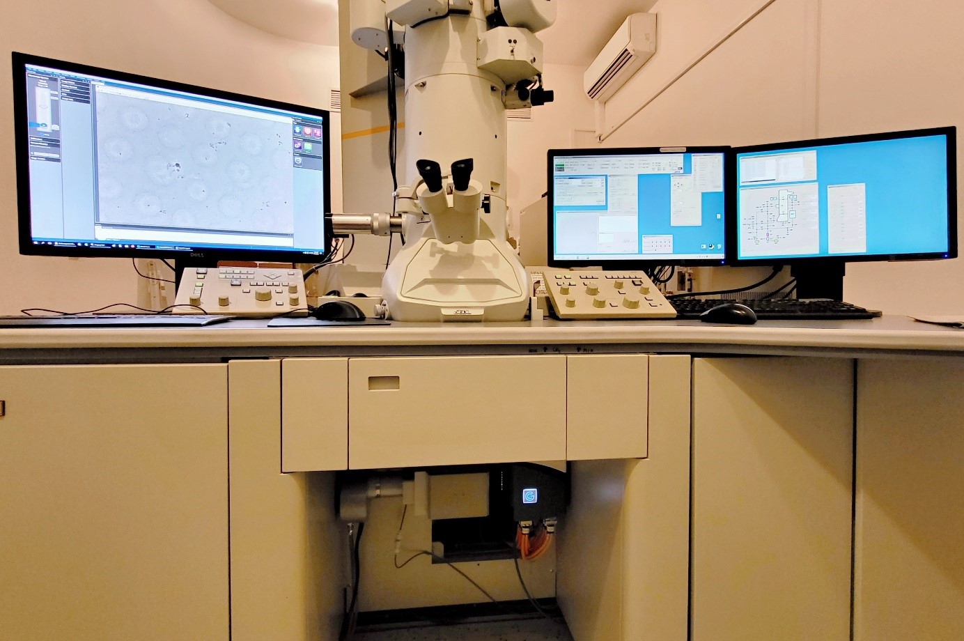
The next level of EM imaging in České Budějovice
Author: Eva Ďurinová
Laboratory of electron microscopy in České Budějovice upgraded their transmission electron microscopes with modern cameras. The new technology allows us to achieve results never thought possible before.
The 200kV microscope JEOL JEM-2100F operates now with K2 Summit™ Direct Detection Camera (Gatan, Inc., USA). Direct Detection cameras detect the incoming electrons directly on the sensor, which removes limitations caused by noise induced by the electron scattering associated with the conversion of primary electrons to photons and the energy deposition processes, which are processes needed by standard/conventional (CCD) TEM digital cameras. The K2 Summit 4k x 4k direct detection camera is also unique in its ability to count individual electrons (instead of integrating the analogue signal as in standard digital cameras). Another benefit of this detector is the super-resolution mode which can pinpoint electron landing coordinates with sub-pixel accuracy and extends the effective resolution to 57 megapixels, allowing a larger specimen area to be imaged at the same resolution while using lower magnification. Also, the sensor read-out speed of 400 full frames per second adds to the great benefits of this detector which is now mainly used for visualization of cryo-samples.
The routine 120kV microscope JEOL JEM-1400 Flash, installed in our laboratory in June 2021, is equipped with a Xarosa bottom-mounted CMOS camera (EMSIS GmbH, Germany). The latest CMOS technology used in this camera brings together high speed (30 frames per second in its full 20-megapixel resolution), high sensitivity and high dynamic range imaging (HDRI). The Xarosa CMOS camera is installed on our Jeol JEM-1400 Flash microscope with RADIUS software which provides several superior features including e.g. drift correction, “click-to-center”, smart averaging, easy image stitching, full interactive measurements and the integrated image database. Although it offers advanced functions, the RADIUS software managed to make the image acquisition super user-friendly. The JEM-1400 microscope equipped by this camera is now used for high resolution imaging of cell ultrastructure at room temperature but is ready to be used for electron tomography or for work in cryo-conditions.
You can follow our facebook for more update from our laboratory @LEMvCB
Focus on technologies
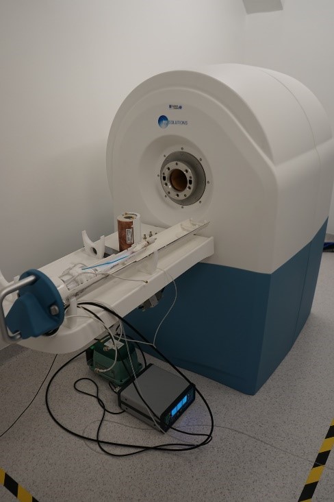
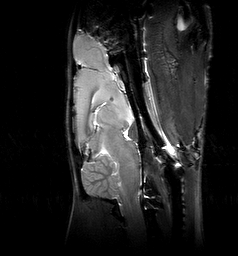
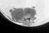
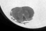
New 7T MRI preclinical scanner in CAPI
Author: Vít Herynek
A new 7T MRI preclinical scanner (MRS*DRYMAG 7.0T, MR Solutions, GB) was installed in CAPI in May 2021. The high-field cryogen free scanner is equipped with radiofrequency coils for scanning of the mouse and rat brain, and mouse and rat body. A set of heated animal beds with monitoring of vital functions and anesthesia makes work with animals easy and comfortable for both the operator and the animals.
 The scanner enables acquisition of highly resolved anatomical
images (proton density, T1- and T2- weighted images) in vivo
virtually of any selected part of rodents’ body, identification of
selected metabolites in the tissue by MR spectroscopy, and
diffusion weighted images.
The scanner enables acquisition of highly resolved anatomical
images (proton density, T1- and T2- weighted images) in vivo
virtually of any selected part of rodents’ body, identification of
selected metabolites in the tissue by MR spectroscopy, and
diffusion weighted images.
Due to the sequence triggering by (breath and/or heart action) the system enables scanning of the abdominal cavity and even the heart in time-resolved image series.
The scanner has a broadband transmit/receive path; therefore, it enables detection also of other nuclei, 19F (using a rat whole body 1H/19F radiofrequency coil) and 31P (1H/31P surface coil).
Output data are both in an intrinsic data format and in DICOM, therefore, they can be easily uploaded and processed by a standard image processing software.


Highlights of user results
A cellular and spatial map of the choroid plexus across brain ventricles and ages
Author: Karol Kaiser
Our group has recently contributed to the publication of a major study in journal Cell - A cellular and spatial map of the choroid plexus across brain ventricles and ages (Dani et al., 2021), published by the groups of prof. Maria Lehtinen and prof. Aviv Regev from Boston Children´s hospital and Broad Institute, respectively.
Main focus of this study was to capture and decipher cellular diversity of the embryonic and adult choroid plexus, which play key role in the production of cerebrospinal fluid throughout life. Using combination of various techniques such as single cell profiling and lineage tracing, this study provides a roadmap for better understanding of age-dependent changes in the tissue physiology and generates novel insights of its developmental trajectory.
In our lab, we focus on the dissection of the role of Wnt signalling pathway in various aspects of vertebrate embryonic development, adult homeostasis as well as in multiple pathological conditions. Recently, we identified novel function of the Wnt signalling in the development of choroid plexus. In this study, we were able to leverage our knowledge of the specific role of the Wnt pathway in the choroid plexus embryogenesis to successfully label and trace fate of cells with active Wnt signaling and reveal their contribution to the developing tissue in vivo. Furthermore, using state-of-the-are in situ hybridization approach, we performed high-resolution transcriptomic profiling of these newly identified cells, thus further clarifying the different aspects of the Wnt pathway activation in the choroid plexus development. In summary, our group helped to confirm the observation from single cell analysis of embryonic choroid plexus and establish novel role of Wnt signaling pathway in the choroid plexus morphogenesis.

Past & upcoming events
Czech‐BioImaging Scientific Conference


The annual Czech BioImaging scientific conference - „Imaging Principles of Life 2022",
will take place 4 – 5 October in the heart of vineyards and almond trees, Hustopeče.
Anyone interested in imaging methods is welcome to enjoy the interesting presentations of expert speakers from
the fields of biological and medical imaging. No presentation is required in order to attend, as the event is
open. The Czech-BioImaging RI users are especially encouraged to participate in this memorable event and join
the Czech-BioImaging family to the fullest. Oral presentations complemented with a poster session will showcase
the most exciting results obtained in Czech-BioImaging facilities. Special space will also be allocated to the
industrial partners for presenting current technology novelties. An integral part of the Conference will, of
course, be a dinner party in the nearby wine cellar První Vinařská, to truly experience the touch of the wine
culture that is so typical for the area.
UPCOMING Educational activities
(spring - summer)
Processing and analysis of microscopic images in biomedicine
Practical course | April 4 - 8, 2022 | IMG, Prague
The course will cover the basics in image data acquisition, processing and analysis including methods of stereology. Apart from a theoretical background, the emphasis will be put on practical experience.
Fundamentals of light microscopy
Course | May 25 - 26, 2022 | CEITEC MU, Brno, CELLIM
Technical aspects of experiments using advanced light microscopy
Course | Spring 2022 | Institute of Physiology CAS, Prague
EM Sample Preparation Series
Practical course | May - June 2022 | UK, BIOCEV, Vestec
Advanced TEM Imaging: From Cells to Proteins
Course | June 13 - 14, 2022 | Biology Centre of CAS, České Budějovice
Super-resolution in light microscopy
Practical course | June 2022 | IMG, Prague
The practical hands-on oriented course is strongly focused on super-resolution methods in light fluorescence microscopy field. The major objective of the course is to introduce newly available methods and cutting-edge machines to potential users: PhD students, postdocs and researchers in the biology.
Upcoming international congresses
16th international congress of histochemistry and cytochemistry
When: 28 – 31 August 2022
Where: Prague
The Society for Histochemistry invite you to Prague in August 2022. The 16th International Congress of Histochemistry and Cytochemistry 2022 brings the worldwide histochemists together to exchange information, closely cooperate and contribute to scientific progress.
REGISTRATION IS NOW OPEN
16th Multinational Congress on Microscopy

When: 4 - 9 September 2022
Where: Brno
Join us at the 16th Multinational Congress on Microscopy and become a part of the unique microscopy ecosystem in Brno. The aim of the 16th MCM is to bring together leading academic scientists, researchers, students and the commercial sphere to exchange and share experiences and research results on all aspects of microscopy.
REGISTRATION IS NOW OPEN
Useful links
Czech-bioimaging Scientific Conference 2022
Czech-bioimaging – pro veřejnost
The National Infrastructure for Biological and Medical Imaging,
Czech-BioImaging, is supported by the Ministry of Education, Youth and
Sports of the Czech Republic (project No. LM2018129) and by European
Regional Development Fund (project No.
CZ.02.1.01/0.0/0.0/18_046/0016045).

