Brno Node – Multimodal Imaging Node Brno
About Brno Node
Location: Brno
Number of Core Facilities: 6
Technologies & expertise:
Medical imaging: Animal ultra-high field MR imaging and spectroscopy (9.4T), potentially combined with focused ultrasound, and human MR imaging (3T) with electrophysiological techniques.
Light microscopy: Imaging of plant systems, mammalian germs cells, stem cells and embryos, and development of image analysis tools, quantitative and rapid measurements of cell behavior, mainly growth and motility
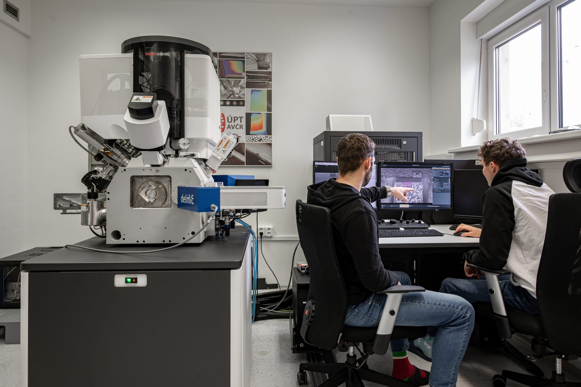
Electron microscopy: Chemical and cryogenic preparation of biological samples, imaging using SEM, cryo-SEM, STEM, FIB-SEM, Raman spectroscopy analysis
Data analysis


Specialties and expertise of the Node
The Medical Imaging part is formed by two closely collaborating facilities. The NMR facility at the Institute of Scientific Instruments specializes in animal ultra-high field MR imaging and spectroscopy (9.4T), possibly in combination with focused ultrasound. The Multimodal and Functional Imaging Laboratory (MAFIL) at CEITEC, Masaryk University, is focused on human MR imaging (3T) accompanied by electrophysiological techniques. Together, these facilities enable translational research and offer a complex portfolio of MRI techniques including multimodal approaches (e.g., simultaneous EEG-fMRI) and human hyper-scanning (fMRI with two participants measured simultaneously in two scanners).
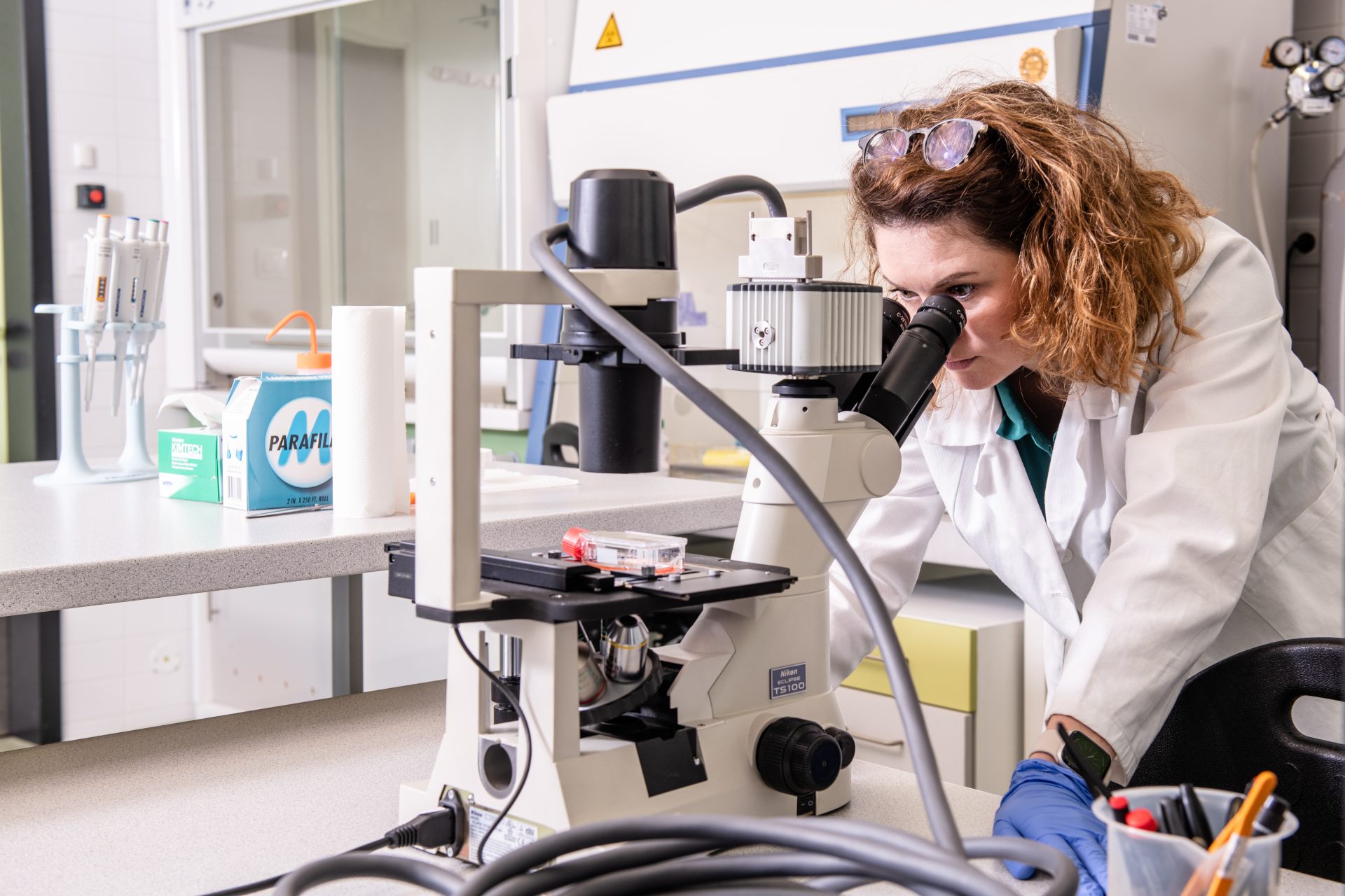
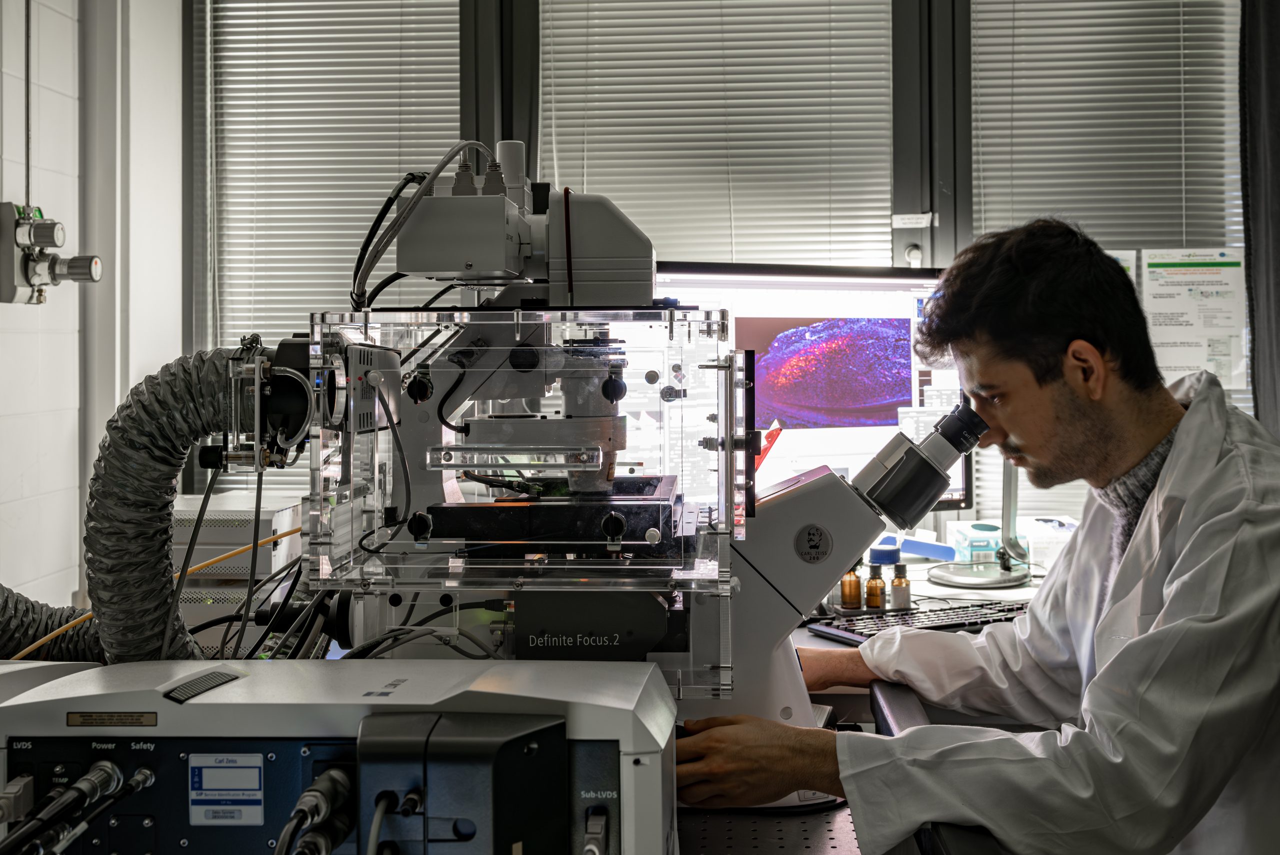
The Microscopy part covers both Advanced Light Microscopy and Electron Microscopy and consists of three individual labs. The core facility Cellular Imaging (CELLIM) at the CEITEC, Masaryk University, is focused on light microscopy, especially in imaging of plant systems, mammalian germs cells, stem cells and embryos, and development of image analysis tools. The Experimental Biophotonics Facility at the CEITEC, Brno University of Technology is focused on highly quantitative and rapid measurements of cell behavior, mainly growth and motility with Q-Phase Multimodal Holographic Microscope. The Core facility Electron microscopy and Raman spectroscopy at the Institute of Scientific Instruments is provides chemical and cryogenic preparation of biological samples, imaging using SEM, cryo-SEM, STEM, FIB-SEM, Raman spectroscopy analysis and also an individual approach in the area of specialized microfluidic techniques.
Centre for Biomedical Image Analysis (CBIA) at the Faculty of Informatics, Masaryk University, offers advanced expertise for the analysis of both biological and medical images and collaborates with the microscopy and medical imaging facilities within the Node.
Instrument highlights
The Brno Node is equipped with two 3T Siemens Prisma scanners for human medical imaging. These scanners are designed specifically for high quality research data based on features like strong gradient fields, excellent homogeneity of mg. field and excitation, high sensitivity with 64 channel head/neck coils. Simultaneous use of two scanners enables a relatively unique feature of hyper-scanning (dual fMRI). The human MR scanners are equipped with several MR compatible electrophysiological devices for recording of high-density EEG, ECG, breathing, skin conductance, etc.
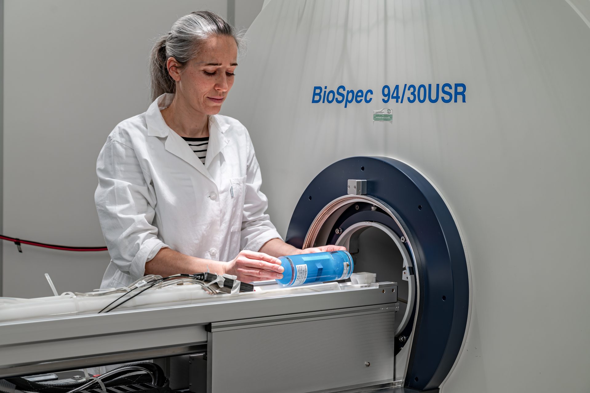
The node is also equipped with a high-field 9.4T MR scanner (Bruker Biospec 94/30), dedicated primarily to preclinical studies involving small laboratory animals (mice, rats and possibly rabbits). Further equipment including a cryo-coil and multi-nuclear RF coils allows advanced MR imaging and spectroscopy examinations. The laboratory also offers a combination of MR with the application of focused ultrasound and services necessary for performing animal experiments (animal facility for 200 mice + 100 rats, specific pathogen free, BSL-1, 1st-category GMO).
Besides providing access to a wide selection of equipment and analysis tools, the light microscopy unit specializes in plant in vivo imaging and techniques useful for research on live mammalian cells, mammalian germ cells, stem cells and embryos. The light microscopy unit CELLIM recently expanded to include a new SIM/SMLM system from Carl Zeiss, the Elyra 7 – Lattice SIM. This instrument provides several imaging modalities, like structural illumination microscopy (SIM), total internal reflection microscopy (TIRF) and single molecule localization microscopy (SMLM), which allows for a wide range of applications.
The Experimental Biophotonics Facility of this Node offers user access to their Q-Phase Multimodal Holographic Microscope developed in-house and commercialized by Tescan / Telight. The microscope provides highly quantitative and rapid measurements of cell behavior, mainly growth and motility, with unprecedented accuracy where the distribution of dry mass inside cells is determined with standard deviation of 0.4 pg/µm2. The microscopy is an implementation of Quantitative Phase Imaging where the dynamic morphometry with live cells in tissue culture is a label free technique and the accuracy is achieved by using an incoherent light source, which also uniquely allows quantitative imaging of cells in 3D environments such as collagen matrix. Integrated fluorescence imaging is available for automated time-lapse with alternating phase imaging and overlaid images are available for examination.
The Core facility Electron microscopy and Raman spectroscopy of the Brno node provides comprehensive user services in sample preparation and access to imaging and analytical methods. These instruments include: SEM Magellan 400/L (Thermo Fisher Scientific) equipped with cryo-stage, EDX detector Octane (EDAX) and CL detector MonoCL4+ (Gatan); DualBeam FIB-SEM Helios (Thermo Fisher Scientific) fitted with STEM detector, CL detector SPARC (Delmic) and in-house developed cryo-stage; Ultra STEM microscope Nion, HERMES™100S; and inVia Renishaw Raman spectrometer for spectral analysis and mapping. Next, there are state-of-the-art devices for sample preparation: High-pressure freezer EM ICE (Leica microsystems); freeze-substitution unit EM AFS2 (Leica microsystems); cryo-vacuum chamber BAF060 (Leica microsystems); cryo-vacuum chamber ACE600 (Leica microsystems); Leica EM CPD300 (Leica microsystems); plunge freezer Leica EM GP2 (Leica microsystems); and ultramicrotome Leica Enuity (Leica microsystems), while also specializing in applications that demand extreme conditions.
The CBIA unit of the Node offers extensive image analysis services, including tailor-made software development, especially for the tasks of object detection, classification, segmentation and tracking. Both standard algorithmic and deep machine learning solutions are designed, implemented, tested and deployed. To facilitate the development of AI-based solutions, the available equipment includes high-performance servers with multiple top GPU cards as well as high-capacity storage space.


Core facilities
- Multimodal and Functional Imaging Laboratory (MAFIL) at CEITEC, Masaryk University
- Magnetic Resonance Core Facility, Institute of Scientific Instruments, Czech Academy of Sciences
- Core Facility Electron Microscopy and Raman spectroscopy, Institute of Scientific Instruments, Czech Academy of Sciences
- Biophotonics Core Facility at CEITEC, Brno University of Technology
- Core Facility Cellular Imaging (CELLIM) at CEITEC, Masaryk University
- Centre for Biomedical Image Analysis (CBIA) at Faculty of Informatics, Masaryk University
Brno Node contacts

Head of the Node & Representative of Medical Imaging
Michal Mikl

Deputy Head of the Node & Representative of Biological Imaging
Milan Ešner


