CAPI Node – Center for Advanced Preclinical Imaging
About CAPI Node
Location: Prague
Number of Core Facilities: 1
Technologies & Expertise:
Primarily in vivo preclinical imaging of small laboratory animals (mice and rats). Besides imaging methods commonly used in clinical practice (MRI, CT, ultrasound, PET, SPECT) the facility uses methods less common (optical imaging) and methods experimental (photoacoustic imaging, magnetic particle imaging). The facility instrumentation thus enables development of new diagnostic procedures, but also solution of specific scientific projects using novel methods and approaches for instance in neurology, cardiology, oncology, theranostics, transplantation and regenerative medicine.


Specialties and expertise of the Node
Examples of research application areas cover cancer research studies on immunocompetent, immunodeficient, and PDX mice, neurology studies including neurodegenerative diseases, functional cardiology studies, vascularization, and also development and characterization of new contrast agents. We are approved for radiation work (open and closed sources of ionizing radiation), and GMO (including GMO-2 cell lines). Our SPF animal facility enables to breed and maintain various transgenic mouse and rat models. We are equipped and skilled for microsurgery, whole-body irradiation (60Co irradiator), stem cell transplantation and tracking, and hematology/immunology characterization.
Our team collaborates with customers on experimental setup, obtaining approvals on experimental animal research, data evaluation and interpretation, and also on publication of results for the given problem in the area of preclinical research.
Selected model applications:
Neurology: study of neurodegenerative diseases (CT, PET, MRI), traumatic injuries (anatomic and diffusion weighted MRI), study of the therapeutic effect of stem cells (MRI), metabolic and functional disorders (MR spectroscopy, functional MRI)
Cardiovascular system: study of heart disorders (e. g., patent foramen ovale) using real-time imaging (ultrasound, photoacoustic imaging), triggered imaging of heart action (MRI), angiography (CT, MRI), 3D and 4D imaging of the heart (high-frequency ultrasound)
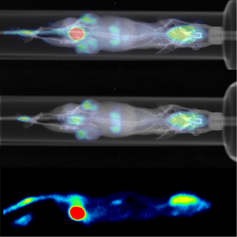
Oncology: monitoring of tumor and metastasis grow (optical imaging, MRI, ultrasound, PET SPECT, CT), monitoring of a therapeutic effect of newly developed cytostatics, targeting of tumor cells by specific contrast agents (optical imaging, MRI, ultrasound/hpohoacoustics, radionuclide methods, volumetry (MRI, ultrasound), tumor oxygenation (photoacoustics), tumor vascularization – MVA (ultrasound – Power/Color Doppler), organ and tumor perfusion (contrast-enhanced MRI, high-frequency ultrasound in a contrast mode using microbubbles)
Transplantation and regenerative medicine: microsurgery, cell microapplication (surgical microscope), monitoring of the transplantation outcome (ultrasound, MRI), cell transplantation and cell tracking (optical imaging, MRI, PET, SPECT)
Contrast and theranostic agent development: cytotoxicity, biocompatibility, biodistribution, verification of specific targeting, possibility of image-guided application of the agent (the smallest bolus ca 3µL) into the tumor, muscle, or a selected organ or tissue (high-frequency ultrasound, syringe pump)
Instrument highlights
1T MRI high throughput machine for basic anatomical screening of mice and rats, possibility of colocalization with PET/SPECT/CT, MPI, OI
7T MRI precise anatomical imaging, MR spectroscopy, diffusion weighted imaging, time-resolved imaging (heart imaging with both prospective and retrospective gating), quantitative imaging (diffusion tensor imaging, relaxometry), X-nuclei imaging and spectroscopy
MPI 3D in vivo imaging (i.e., imaging of distribution of the injected magnetic tracer) with 20-ms temporal resolution, possibility to quantify the tracer amount. Suitable for tumor imaging and in situ, hyperthermia therapy (MFH), acute stroke detection, molecular imaging, stem cell tracking, characterization of magnetic nanoparticles, targeted tissue imaging.
US & PA or multimodal imaging in oncology (tumor detection & sizing in 2D + 3D, vascularization, hypoxia), molecular biology (characterization of nanoparticles, dyes and contrast agents; drug delivery and pharmacokinetic, cell tracking), neurobiology (oxygen saturation, total hemoglobin and blood flow velocity), cardiology (cardiac function in 2D, 3D and 4D, ischemia & hypoxia).
Available transducers are:
- MX201 (10-22MHz, Centre Transmit: 15 MHz) – mouse whole body imaging, brain (mouse, rat), cardiovascular and abdominal imaging, tumor imaging, neurobiology
- MX400 (20-46 MHz, Centre Transmit: 30 MHz) – mouse abdominal imaging, vascular imaging, embryology, lymph nodes, tumor imaging
- MX550D (25-55 MHz, Centre Transmit: 40 MHz) – mouse & Rat eye imaging, skin, abdominal & microvasculature imaging, lymphatics & reproductive imaging MX700 (29-71 MHz, Centre Transmit: 50 MHz) – vascular, embryology, superficial tissue, ophthalmology imaging


Core facility
CAPI contacts

Head of the Node
Luděk Šefc

Project coordinator
Pavla Volešák Francová


